Home » Without Label » Anatomical Name Of Lower Back Muscles : Muscles Move And Support The Spine / The quadratus lumborum muscles (orange, in the image above) are found in the lower back (also called the lumbar area).
Anatomical Name Of Lower Back Muscles : Muscles Move And Support The Spine / The quadratus lumborum muscles (orange, in the image above) are found in the lower back (also called the lumbar area).
Anatomical Name Of Lower Back Muscles : Muscles Move And Support The Spine / The quadratus lumborum muscles (orange, in the image above) are found in the lower back (also called the lumbar area).. Muscles of ulnar nerv 12 photos of the muscles of ulnar nerv muscles of ulnar nerve, muscles the ulnar nerve stimulates, muscular branches of ulnar nerve, human muscles, muscles of ulnar nerve, muscles the ulnar nerve stimulates, muscular branches of ulnar nerve The quadratus lumborum muscles (orange, in the image above) are found in the lower back (also called the lumbar area). The superficial group, also known as the appendicular group, is primarily associated with movement of the appendicular skeleton. The lumbar spine is the lower back that begins below the last thoracic vertebra (t12) and ends at the top of the sacral spine, or sacrum (s1). These muscles provide posture and stability to the body by holding the vertebral column erect and adjusting the position of the body to maintain balance.
1 your spine in this region has a natural inward curve. Anatomical name of lower back muscles : The trapezius or trapezoid muscles are two paired muscles that extend from the base of the thoracic vertebrae in the spine to the occipital bone and run out to the spine of the scapula. In turn, the posterior deep muscles are the piriformis, obturator internus, obturator externus, superior gemellus, inferior gemellus, and quadratus femoris. Most of the time, back muscle pain is diagnosed then treated with little more than a prescription of rest, painkillers and muscle relaxants.
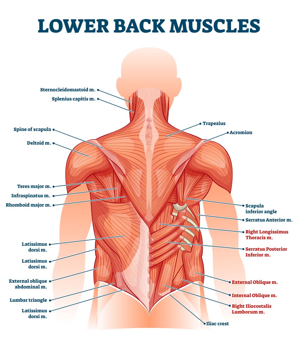
Lower Back Muscle Anatomy And Low Back Pain from ix-cdn.b2e5.com The quick answer to this question is the muscles of the lower back are the multifidus, longissimus, spinalis, and quadratus lumborum. Anatomical name of lower back muscles : The muscles of the back with the surface (trapezius, latissimus dorsi, thoracolumbar fascia, deltoid) and intermediate layers (serrated muscles, external and internal oblique muscle). The lordotic curve your lower back (lumbar spine) is the anatomic region between your lowest rib and the upper part of the buttock. The pelvic floor muscles also help increase this pressure, which provides stability to the spine and trunk. This blog post article is an overview of the muscles of the lumbar spine of the trunk. As you can see, there are also have a spine of scapula deltoid, triceps brachii, latissimus dorsi. The muscles that move the upper legs (thigh) there are many muscles that move the large bone of the thigh.
The muscle then courses up to your shoulder and attaches to your upper arm bone.
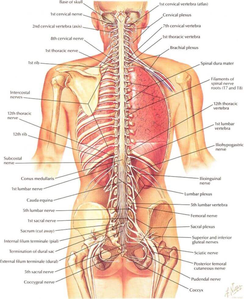
See back muscle anatomy stock video clips. When it contracts, it makes the foot bend downward, and it also helps to bend the knee. 1 your spine in this region has a natural inward curve. It is innervated by anterior rami of spinal nerves, reflecting its embryological origin outside the back. The muscle then courses up to your shoulder and attaches to your upper arm bone. The back anatomy includes the latissimus dorsi, trapezius, erector spinae, rhomboid, and the teres major. The muscles that move the upper legs (thigh) there are many muscles that move the large bone of the thigh. In this image, you will find an occipital bone, sternocleidomastoid, trapezius, deltoid in muscles of the lower back diagram. The vertebral column of the lower back includes the five lumbar vertebrae, the sacrum, and the coccyx. The superficial back muscles include the suboccipital muscles, trapezius, latissimus dorsi, levator scapulae, rhomboids and serratus posterior muscles. These structures work together to support the body, enable a range of movements, and send messages from the brain to. In turn, the posterior deep muscles are the piriformis, obturator internus, obturator externus, superior gemellus, inferior gemellus, and quadratus femoris. On this page, you'll learn about each of these muscles, their locations and functional anatomy.
Muscles of the lumbar spine. The vertebral column of the lower back includes the five lumbar vertebrae, the sacrum, and the coccyx. They originate from the thoracolumbar fascia, the spinous process of thoracic six through 12, the iliac crest, and your lower three ribs. The trapezius or trapezoid muscles are two paired muscles that extend from the base of the thoracic vertebrae in the spine to the occipital bone and run out to the spine of the scapula. Anatomical name of lower back muscles :
Hamstring Muscles And Your Back Pain from www.verywellhealth.com This depth, combined with the fact that the psoas originates from the sides of the five lumbar vertebrae, means it plays an important role in back health. The lower part of the trapezius ascends and depresses the scapula, while the transverse or middle region of the trapezius is what retracts the. When it contracts, it makes the foot bend downward, and it also helps to bend the knee. Muscles of the lumbar spine. They help to bend the back to one side or the other. In this image, you will find an occipital bone, sternocleidomastoid, trapezius, deltoid in muscles of the lower back diagram. The trapezius or trapezoid muscles are two paired muscles that extend from the base of the thoracic vertebrae in the spine to the occipital bone and run out to the spine of the scapula. Let us introduce you to each of these muscles presented in our diagram.
(2017, elsevier) should be consulted.
Your lats are a major back muscle and mover of your shoulder joint. The lordotic curve your lower back (lumbar spine) is the anatomic region between your lowest rib and the upper part of the buttock. These muscles include the large paired muscles in the lower back, called erector spinae, which help hold up the spine, and gluteal muscles. This picture also contains humerus, olecranon process of ulna, deep to tendon and so on. The superficial back muscles include the suboccipital muscles, trapezius, latissimus dorsi, levator scapulae, rhomboids and serratus posterior muscles. The anatomy of the back refers to the muscles of the back, as well as the bones of the scapulae, ribcage, and spine.covering an expanse from the neck to the tailbone, the back muscles are responsible for a broad range of functions, from extending the spine to shrugging the shoulders. These structures work together to support the body, enable a range of movements, and send messages from the brain to. 1 your spine in this region has a natural inward curve. These bones work together to provide flexibility to the trunk, support the muscles of the trunk, and protect the spinal cord and spinal nerves of the back. This depth, combined with the fact that the psoas originates from the sides of the five lumbar vertebrae, means it plays an important role in back health. Anatomical name of lower back muscles : To perform clinical clinical orthopedic manual therapy to the lumbar spine. The quick answer to this question is the muscles of the lower back are the multifidus, longissimus, spinalis, and quadratus lumborum.
This muscle is the largest flexor of the foot. This curve, called lordosis, helps to: The lower part of the trapezius ascends and depresses the scapula, while the transverse or middle region of the trapezius is what retracts the. The lumbar spine is the lower back that begins below the last thoracic vertebra (t12) and ends at the top of the sacral spine, or sacrum (s1). The psoas muscle is a low back muscle located deep in the body, very close to the spine and inside the hip and thigh bones.
The 7 Best Lower Back Stretches For Pain Legion from legionathletics.com The quadratus lumborum muscles (orange, in the image above) are found in the lower back (also called the lumbar area). Its name means belly of the leg,and its common name is the calf muscle. Let us introduce you to each of these muscles presented in our diagram. Anatomical name of lower back muscles : The superficial group, also known as the appendicular group, is primarily associated with movement of the appendicular skeleton. In turn, the posterior deep muscles are the piriformis, obturator internus, obturator externus, superior gemellus, inferior gemellus, and quadratus femoris. The muscles of the back with the surface (trapezius, latissimus dorsi, thoracolumbar fascia, deltoid) and intermediate layers (serrated muscles, external and internal oblique muscle). These muscles include the large paired muscles in the lower back, called erector spinae, which help hold up the spine, and gluteal muscles.
Most of the time, back muscle pain is diagnosed then treated with little more than a prescription of rest, painkillers and muscle relaxants.
The back muscles represented on an anatomical chart and on a schematic view of the origin and insertion of the proper muscles of the back (iliocostal muscle of. In this image, you will find an occipital bone, sternocleidomastoid, trapezius, deltoid in muscles of the lower back diagram. This blog post article is an overview of the muscles of the lumbar spine of the trunk. They help to bend the back to one side or the other. The lordotic curve your lower back (lumbar spine) is the anatomic region between your lowest rib and the upper part of the buttock. It is composed of trapezius, latissimus dorsi, rhomboid major, rhomboid minor and levator scapulae. The muscle then courses up to your shoulder and attaches to your upper arm bone. The back anatomy includes the latissimus dorsi, trapezius, erector spinae, rhomboid, and the teres major. Anatomical name of lower back muscles : The lower part of the trapezius ascends and depresses the scapula, while the transverse or middle region of the trapezius is what retracts the. When it contracts, it makes the foot bend downward, and it also helps to bend the knee. In turn, the posterior deep muscles are the piriformis, obturator internus, obturator externus, superior gemellus, inferior gemellus, and quadratus femoris. The back consists of the spine, spinal cord, muscles, ligaments, and nerves.
:max_bytes(150000):strip_icc()/Depositphotos_19871399_original-56a05f523df78cafdaa14cd1.jpg)